The experimental apparatus consists of an x-ray spectroscopy end station of moderate energy resolution operating with proton-induced quasi-monochromatic x-ray beams. It was designed, installed and operated at the 5.5 MV Tandem Accelerator of the INPP with the support of several funded projects (PEP Attikis Framework, Project ATT-29, 2006 –2008, DEMO-RESEARCH, 2007-2008 and of an IAEA Coordinated Research Project, No. 13833, 2006-2009). The apparatus includes a two-level ultrahigh vacuum chamber that hosts in the lower level up to six primary targets in a rotatable holder; there, the irradiation of pure element materials—used as primary targets—with few-MeV high current (∼μA) proton beams produces intense quasi-monochromatic x-ray beams of selectable energy within the 1-10 keV regime. In the chamber’s upper level, a six-position rotatable sample holder hosts the targets considered for x-ray spectroscopy studies. The proton-induced x-ray beam, after proper collimation, is guided to the sample position whereas various filters can be also inserted along the beam’s path to eliminate the backscattered protons or/and to absorb selectively components of the x-ray beam. The apparatus incorporates an ultrathin window Si(Li) spectrometer (FWHM 136 eV at 5.89 keV) coupled with low-noise electronics capable of efficiently detecting photons down to carbon Kα.
The apparatus optimized experimental conditions have been so far utilized to study specific atomic physics related topics such as resonant Raman scattering and the soft x-ray cascade emission demonstrating that this methodology offers for these types of studies. Its application for selective XRF analysis using multiple excitation energies has been also shown to exhibit important advantages for a reliable quantification along a rather extended energy range, as well as to significantly improve the detection limits for low-Z elements. Future applications, envisage the application of the apparatus to produce intense soft X-ray beams within the ‘water window’ for structural analysis of biological specimen and the study of dual (charged particle and X-ray beams) inner shell ionization of samples relevant for modelling and validating quantitative procedures of analysis to be employed for passive spectrometers being developed for planetary missions.
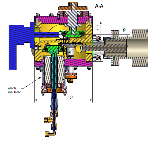
Transverse cross section of the PIXE-XRF scattering chamber
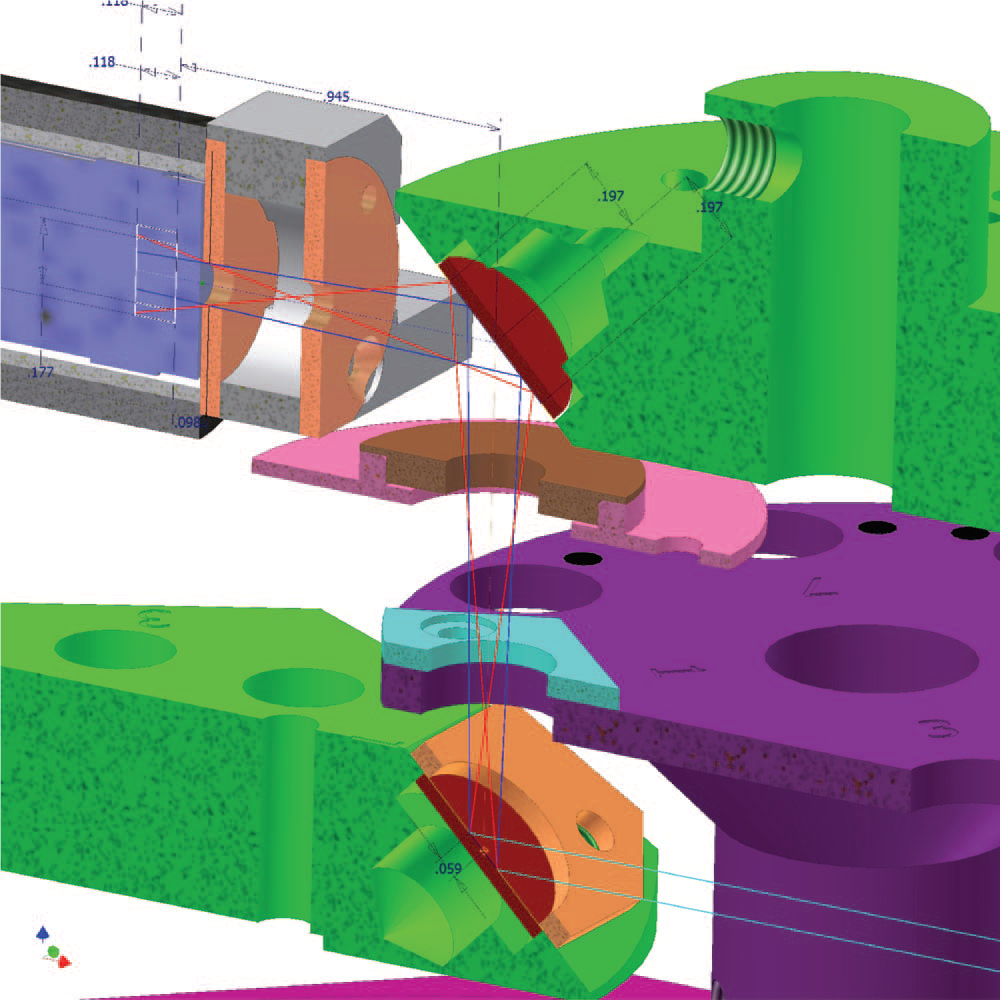
Schematic drawing of the PIXE-XRF scattering chamber allowing multiple electrically isolated and water-cooled primary targets and samples to be placed within an optimized geometrical configuration
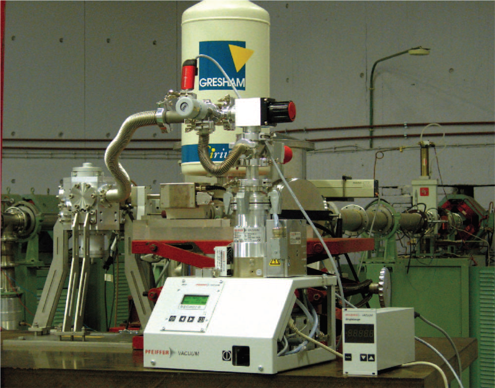
The experimental Apparatus for the production of Particle Induced Monoenergetic X-ray Beams (PIXE-XRF) installed at the TANDEM beamline